Varus deformity of the hip. Varus deformity of the femoral neck
The deformity of the femoral neck is based on a decrease in the cervical-diaphyseal angle and shortening of the neck. The main manifestations are duck gait, lumbar lordosis, limited rotation and abduction of the hip in the hip joint. it is symptomatic, juvenile and childish, congenital isolated, which is quite rare.
With congenital isolated deformity of the neck, the newborn has a high location of the greater trochanter of the femur and shortening of the limb. The absence of ossification nuclei makes the diagnosis difficult. After the onset of ossification, a shortening of the femoral shaft, a bent neck, and adduction of the distal end of the femur are detected. The greater trochanter stands high and rebuilt in a coracoid shape, the acetabulum flattens, the femoral head shifts backwards and downwards, the epiphyseal germ zone is located vertically.
At the age of three to five years, children's varus deformity develops, which is manifested by the formation of a trihedral bone fragment in the lower medial part of the femoral neck. At the same time, enlightenments are formed in the area of the neck and head. The bony edges at the fissure are uneven, serrated, slightly sclerotic, the course of the fissure is tortuous. Over time, the gap can expand up to ten to twelve millimeters, the development of the head lags behind, it shifts caudally, approaching the femoral shaft, and the neck shortens.
Juvenile varus deformity of the neck is characterized by changes in the growth zone. Enchondral growth zone expands early due to resorption bone tissue loosened. Gradually and slowly, the femoral head slides down, inwards and backwards. Epiphysiolysis of the femoral head develops. Pathological processes in the upper metadiaphysis or femoral neck cause symptomatic varus deformity. The opposite of varus deformity is valgus deformity, which can be acquired or congenital.
Among orthopedic diseases, congenital hip dislocation accounts for three percent, and hip dysplasia occurs in sixteen cases per thousand births. Hip dislocation can be unilateral or bilateral. The cause of dislocation is hip dysplasia, which affects all components of the joint: flattening and hypoplasia of the acetabulum; slowing down ossification and hypoplasia of the femoral head; anomalies in the development of the neuromuscular apparatus.
The head is in an eccentric position, and the acetabulum is underdeveloped with congenital subluxation. In the future, congenital dislocation develops, the main clinical signs of which are: shortening of the lower limb, asymmetry of the gluteal folds, limitation of hip abduction, slipping symptom, gait disturbance (when the child begins to walk). The main radiographic findings are: a vertical line (if there are no ossification nuclei) that passes through the outer superior acetabular ridge, crossing the inner edge of the thigh. In this case, the presence of a broken line of Calvet and a ledge-like line of Menard - Shenton is characteristic. In addition to these symptoms, there are antetorsion of the neck, thickening and shortening of the femoral neck, deformity of the head, bone atrophy on the side of dislocation, etc.
The invention relates to medicine, namely to orthopedics, traumatology in the treatment of varus deformity of the femoral neck. Essence: wires are passed through the iliac wing, greater trochanter, middle and lower thirds of the thigh, the ends of the wires are fixed on the supports of the compression-distraction apparatus, the support on the wing of the ilium and the proximal support on the thigh are connected, and the middle support is connected to the distal one on the thigh, perform intertrochanteric osteotomy of the femur in the direction from the bottom up, from the outside - inside, the deformity of the proximal femur is corrected, a transverse osteotomy is performed in the lower third of the femur, the intermediate fragment of the femur is shifted medially, fixed in the achieved position, cantilevered wires are passed through the greater trochanter and the femoral neck, needles are passed through the supraacetabular region, they are bent in an arcuate manner, fixed and pulled to the arc of the apparatus, on the 5-6th day after the operation, distraction is carried out between the middle and distal supports at a faster pace along the outer rods of the apparatus, which allows forming the roof of the acetabulum, leveling the length of the limb, but rmalize the biomechanical axis. 5 ill.
The invention relates to medicine, in particular to orthopedics and traumatology, and in particular is used in the treatment of varus deformity of the femoral neck using a transosseous fixation apparatus A known method for the reconstruction of the hip joint, providing for the simultaneous restoration of the cervical-diaphyseal angle (NDA) and an increase in the coverage of the femoral head by supraacetabular osteotomy of the ilium and tilting the distal fragment of the pelvis outward (AS 757155, USSR. A method for correcting the cervical-diaphyseal angle and the acetabular roof cavities in varus deformity of the femoral neck, published April 28, 1980, Bull. 31). However, this method involves performing a subtrochanteric wedge-shaped or intertrochanteric angled osteotomy, supraacetabular osteotomy, followed by fixation with a plaster cast, which does not allow gently forming the roof of the acetabulum, eliminating the pathological restructuring of the femoral neck, completely equalizing the length of the limb and normalizing its biomechanical axis. The objective of the present invention is to develop a method for the treatment of varus deformity of the femoral neck, which allows to increase the coverage of the femoral head without osteotomy of the ilium, eliminate the pathological restructuring of the femoral neck, completely equalize the length of the limb and normalize its biomechanical axis. The problem is solved by the fact that in a method for treating varus deformity of the femoral neck, including performing intertrochanteric osteotomy and fixing fragments of the femur and ilium in the supports of the transosseous apparatus, additionally injected through the region of the greater trochanter, the femoral neck, at least four cantilevered wires, and through the supraacetabular region - at least two wires, the ends of which are bent outwards, fixed in the support of the apparatus and pulled, while in the lower third, transverse osteotomy of the femur is performed, and intertrochanteric osteotomy is performed in the direction from bottom to top from the outside inward, after which the intermediate fragment is moved under the zone of pathological restructuring of the neck hips. The present invention is explained detailed description , clinical example, scheme and photographs in which: Fig. 1 depicts a diagram of osteotomy of the femur with fixation of its fragments and the hip joint in the supports of the transosseous apparatus; figure 2 is a photo of the patient E. before treatment; figure 3 shows a copy of the R-gram of the patient E. before treatment; figure 4 illustrates a photo of the patient E. after treatment; figure 5 is a copy of the R-gram of the patient E. after treatment. The method is carried out as follows. In the operating room after anesthesia treatment of the surgical field with an antiseptic solution, the needles are carried out at four levels (figure 1): through the wing of the ilium, the region of the greater trochanter, the middle and lower thirds of the thigh. The ends of the wires passed through the bone are fixed in pairs on the supports of the compression-distraction apparatus. The support on the wing of the ilium and the proximal support on the thigh are connected to each other by means of hinges; the middle support and the distal one on the thigh are connected to each other using threaded rods. The connected supports are movable relative to each other. Then perform intertrochanteric osteotomy of the femur in the direction from the bottom up from the outside - inside. The deformity of the proximal femur is corrected. In the lower third of the thigh, its transverse osteotomy is performed and the medial shift of the intermediate fragment of the femur is performed. After that, the fragments of the femur are fixed with the help of supports in the achieved position. Cantilever wires are passed through the greater trochanter and femoral neck, and wires are passed through the supraacetabular region, which are arcuately bent, fixed and pulled to the arc of the transosseous fixation apparatus, which contributes to the stimulation of reparative processes in the femoral neck and acetabular roof. On the 5th-6th day after the operation, distraction is carried out between the middle and distal femoral supports at a faster rate along the outer rods of the apparatus, while forming a trapezoidal regenerate until the length of the limbs is equalized with the restoration of its biomechanical axis. After achieving complete consolidation in the areas of osteotomy, the apparatus is dismantled. An example of the implementation of the method. Patient E. (case history 30556) was admitted for treatment with the following diagnosis: Consequences of hematogenous osteomyelitis, varus deformity of the neck of the right femur - 90 o , shortening of the right lower limb 4 cm, combined contracture of the right hip joint (extension - 160 o , abduction - 100 o), valgus deformity of the knee joint - 165 o . The duration of the disease is 5 years (figure 2). Upon admission, he complained of fatigue, recurrent pain in the right hip joint, lameness, shortening of the right lower limb, limitation of movement in the right hip joint, and deformity of the right lower limb. Trendelenburg's symptom is sharply positive. On the radiograph of the pelvis - deformity of the proximal femur, SDA - 90 o . Destruction of the femoral neck with its fragmentation throughout is noted. The acetabulum is dysplastic: the acetabular index (AI) is 32 o , the thickness index of the bottom of the acetabulum (ITDV) is 1.75, the depth index is 0.3. In the operating room, after anesthesia, the treatment of the surgical field with an antiseptic solution, wires were inserted at four levels: through the iliac wing, the region of the greater trochanter, the middle and lower thirds of the thigh. The ends of the wires passed through the bone are fixed on the supports of the compression-distraction apparatus. The support on the wing of the ilium and the proximal support on the thigh are connected to each other by means of hinges; the middle support and the distal one on the thigh are connected to each other by means of threaded rods. Then intertrochanteric osteotomy of the femur was performed in the direction from the outside - inside from the bottom up and transverse osteotomy in the lower third of the thigh. The deformity of the proximal femur was corrected and the intermediate fragment of the femur was shifted medially. After that, the fragments of the femur are fixed with the help of supports in the achieved position. Cantilever wires are passed through the greater trochanter and femoral neck, and through the supraacetabular region - wires that are arcuately curved, fixed and stretched to the arc of the transosseous fixation apparatus. On the 5th-6th day after the operation, distraction was carried out between the middle and distal femoral supports at a faster rate along the outer rods of the apparatus until the length of the limbs was equalized and its biomechanical axis was restored, while a trapezoidal regenerate was formed. The distraction was 27 days. The apparatus was removed after 76 days. After treatment, there are no complaints, the gait is correct, the length of the legs is the same, the Trendelenburg symptom is negative, the range of motion in the hip and knee joints is complete (figure 4). On the radiograph of the pelvis, the centering of the femoral head in the acetabulum is satisfactory, SDU - 125 o , AI-21 o , ITDI - 2.3, the index of the depth of the acetabulum - 0.4 (figure 5). The proposed method of treatment is used in the clinic of the RRC "VTO" them. Academician G.A. Ilizarov in the treatment of patients with varus deformity of the femoral neck. The implementation of this method allows to achieve good anatomical and functional results by eliminating the deformity of the proximal femur, restoring the integrity of the femoral neck, sparing formation of the acetabular roof by stimulating reparative processes by additionally introduced wires into the femoral neck and acetabular roof, restoring the biomechanical axis of the limb when simultaneous unloading of the hip joint with a transosseous fixation device. The proposed method involves the use of well-known tools produced by the medical industry, does not require additional accessories, devices, expensive materials and is relatively low-impact. Allows functional load on the operated limb and exercise therapy in the early postoperative period, which prevents the development of persistent contractures of adjacent joints.
Claim
A method for treating varus deformity of the femoral neck, including performing an intertrochanteric osteotomy and fixing the fragments, characterized in that the spokes are passed through the iliac wing, the greater trochanter, the middle and lower thirds of the thigh, the ends of the spokes are fixed on the supports of the compression-distraction apparatus, the support is connected to the wing iliac bone and proximal femoral support, middle femoral support with the distal one, intertrochanteric osteotomy of the femur is performed in the direction from the bottom up, from the outside - inwards, the deformity of the proximal femur is corrected, transverse osteotomy is performed in the lower third of the femur, the intermediate fragment of the femur is shifted medially, fixed in the position reached, cantilevered wires are passed through the greater trochanter and the femoral neck, the needles are passed through the supraacetabular region, they are bent in an arcuate manner, fixed and pulled to the apparatus arc, on days 5-6 after the operation, distraction is carried out between the middle and distal supports with advanced m pace along the outer rods of the apparatus.
Varus deformity of the femoral neck- the basis is the shortening of the neck and a decrease in the cervical-diaphyseal angle. Manifested by limited abduction and rotation of the hip in the hip joint, lumbar lordosis and duck gait. Radiologically, the following deformities are distinguished: congenital isolated, childhood, youthful and symptomatic. Congenital varus deformity is rare.
At newborn shortening of the limb is determined, the greater trochanter of the thigh is located high. In the absence of ossification nuclei, it is difficult to make a diagnosis. When ossification occurs, then a bent neck and shortening of the femoral shaft are found. The distal end of the thigh is shown. The epiphyseal growth zone is located vertically, the femoral head is displaced downwards and backwards, the acetabular cavity is flattened, the greater trochanter is beak-shaped and stands high.
With pediatric varus deformations, which develops at the age of 3-5 years, in the lower medial part of the femoral neck, laterally from the zone of the growth cartilage, a trihedral bone fragment is formed, forming with the upper vertical zone of enlightenment in the region of the head and neck, a picture similar to an inverted letter "U". The course of the fissure is usually tortuous, the bony edges are jagged, uneven, slightly sclerotic.
Later on, the gap expands up to 10-12 mm, the neck is shortened, the head lags behind in development, shifts caudally and approaches the femoral shaft, the greater trochanter is located 4-5 cm above the upper edge of the acetabulum.
juvenile varus deformation is characterized by changes in the growth zone, and not in the bone part of the neck, as in the children's form. In the early stage, the endochondral growth zone expands, loosens due to resorption of bone tissue. In the future, the femoral head begins to slowly and gradually slide down, inwards and backwards, i.e., epiphysiolysis of the femoral head develops.
Symptomatic varus deformity is caused by a pathological process of the femoral neck or its upper metadiaphysis.
Valgus deformity proximal end of the thigh- deformity opposite to varus. It is congenital and acquired. If normally the cervical-diaphyseal angle ranges from 115-140°, then with valgus deformity it can approach 180°, then the axis of the femoral shaft serves as a direct continuation of the axis of the neck.
Congenital dislocation of the hip- population frequency 0.2-0.5%. It accounts for 3% of orthopedic diseases. The frequency of hip dysplasia is 16 cases per 1000 births. The dislocation is unilateral and bilateral in 20-25% of cases. The basis for the occurrence of dislocation is hip dysplasia, affecting all its components: the acetabulum (hypoplasia, flattening), the femoral head (hypoplasia, slowing down ossification), the neuromuscular apparatus (developmental anomalies).
At congenital subluxation of the acetabulum underdeveloped, the head occupies an eccentric position. Then a congenital dislocation develops. The main clinical signs are: a symptom of slipping - a symptom of Marx (a symptom of instability, a click), limitation of hip abduction, asymmetry of the gluteal folds, shortening of the lower limb, and with the beginning of the child's walking - gait disturbance.
Main radiological symptoms: in the absence of ossification nuclei, a vertical line passing through the upper outer protrusion of the acetabulum crosses the inner edge of the so-called femoral beak, which is more distant from the ischium than on the healthy side; the index of the acetabulum (acetabular index) reaches 35-40°; the ledge-like line of Menard - Shenton and the dashed line of Calvet are characteristic; the distance from the most protruding proximal surface of the thigh to the Hilgenreiner line (acetabular line connecting both Y-shaped cartilages) is less than 1 cm.
In the presence of nuclei ossification in addition to these symptoms, the following are revealed: the Hilgenreiner line crosses the head or is located below it; ossification on the side of the dislocation is delayed, the point of ossification of the head is smaller, the sciatic-pubic synchondrosis is open more widely, on the side of the dislocation there is atrophy of the bones, deformity of the head, shortening and thickening of the femoral neck, antetorsion of the neck. The horizontal line drawn along the lower edge of the femoral neck passes above the so-called teardrop, or Kohler's comma, the Maykova-Stroganova symptom is characteristic - the “crescent figure” is superimposed on the medial contour of the femoral neck, etc.
 Varus deformity of the femoral neck Cervical-diaphyseal angle less than average (120 -130°) Causes: § Congenital dislocation of the hip § Juvenile epiphysiolysis § traumatic § rachitic deformity § in case of systemic diseases: fibrous osteodysplasia, pathological bone fragility, dyschondroplasia § consequence of surgical interventions in the area femoral neck § consequences of osteomyelitis, tuberculosis, subcapital osteochondropathy
 Clinic: Congenital - duck gait fatigue in the hip joint during walking. functional shortening of the limb by 3-5 cm or more; limitation of abduction in the hip joint; positive Trendelenburg symptom. Treatment: Subtrochanteric osteotomy
 Valgus deformity of the femoral neck Increase in the neck-diaphyseal angle. ü Congenital ü Traumatic ü Paralytic Clinic: no visible deformities § With concomitant deformities of the knee and foot, gait changes, cosmetic defects Treatment: 1) exercises and corrective postures (“in Turkish”) 2) operative: subtrochanteric osteotomy of the femur.
 Varus and valgus deformity of the knee joints Causes: § congenital, § rickets, § early rising to the feet Varus deformity - the angle is open inwards, Onogi Valgus deformity - the angle is open outwards, X-legs

 Valgus deformity Varus deformity increase in the external condyle, decrease in the internal - compression of the internal meniscus increase in the internal condyle, decrease in the external - compression of the external meniscus the joint space is wider on the outside the joint space is wider on the inside the ligaments are stretched, strengthening the knee joint from the later. the sides are stretched ligaments that strengthen the knee joint on the medial side of the lower leg are often curved with a bulge outward, flat-varus foot setting (clubfoot) flat-valgus foot setting (flat feet) in severe cases: rotation (turn) of the thigh outward, and the lower leg (its lower third) inwards. v Unilateral v Bilateral: symmetrical (concordant deformity) / discordant deformity.
 Diagnosis 1) Goniometer 2) Distance m/d medial. ankles (exceeds 1.5-2.0 cm - up to 2 years, 3 cm - 3-4 years and 4 cm - older) 3) X-ray - 3 degrees
 Treatment 1) 2) 3) 4) Massage Therapeutic gymnastics Orthopedic shoes Surgical treatment- valgus and varus osteotomy


 Flat feet - a change in the shape of the foot, characterized by the omission of its longitudinal and transverse arches. TYPES: longitudinal flatfoot transverse flatfoot longitudinal-transverse
 Foot arches Longitudinal arches: 1) External / cargo (calcaneal, cuboid, IV and V metatarsal bones) 2) Internal / spring (talar, navicular and I, III metatarsal bones) Transverse arch (metatarsal bones heads)
 Etiology Acquired Rachitic platypodia Paralytic platypodia (AFTER POLIO) Traumatic platypodia (ANKLE, CANEAL, AND TARSAL FRACTURES) Static platypodia (excessive load on the foot) Congenital
 Clinic Complaints: § fatigue, pain in the calf muscles by the end of the day § pain in the arch of the foot when standing and walking Typical signs: Ø lengthening of the foot and expansion of its middle section Ø decrease or complete disappearance of the longitudinal arch (the foot rests on its entire plantar surface) Ø abduction (valgus abduction) of the forefoot (toe looks outward) Ø pronation (outward deviation) of the calcaneus over 5 -6 °; In this case, the inner ankle protrudes, and the outer one is smoothed.

 Stages of flat feet q. Hidden stage q. Stage of intermittent flat feet q. Stage of development of a flat foot q. Stage of flat-valgus foot q. Contracture flatfoot

 Diagnosis 2) Podometry according to Friedland - determination of the percentage ratio of the height of the foot and its length (N = 31 -29) 3) Line of Face - a line drawn from the top of the inner ankle to the lower surface of the base of the head of the I metatarsal bone (in N- does not cross the top of the navicular bone )
 Diagnosis 4) Clinical method (normal vault 55-60 mm, angle 90◦) 5) X-ray method (normal vault 120-130◦, normal vault 35 mm)
 Flatfoot degree I degree: Friedland index 25 - 27 clinical angle 105◦ radiological angle up to 140◦ arch height less than 35 mm II degree: radiological angle up to 150◦ arch height less than 25 mm signs of def. Osteoarthritis III degree: radiographic angle up to 170 -175 ◦ arch height less than 17 mm flatness of the forefoot
 Treatment q In the stage of development of a flat foot: I degree: warm foot baths, massage, exercise therapy to strengthen the muscles of the lower leg, wearing arch support insoles II degree: + wearing orthopedic shoes III degree: + surgical treatment
 q In the stage of contracture flat feet Non-operative: blockade of the posterior tibial nerve; ü plaster bandages Operative: ü three-articular arthrodesis of the foot (talonavicular, calcaneocuboid, subtalar) After the onset of arthrodesis, it is necessary to wear orthopedic shoes to form the arch of the foot
 ü Operation according to F. R. Bogdanov - resection of the calcaneocuboid and talo-navicular joints with subsequent arthrodesis of these joints in the corrected position of the arches - lengthening of the tendon of the short peroneal muscle - transplantation of the tendon of the long peroneal muscle on the inner surface of the foot - lengthening of the calcaneal tendon with the elimination of pronation heel and abduction of the forefoot ü Kuslik M. I. operation - crescent-transverse resection of the foot - lengthening of the calcaneal tendon - transplantation of the tendon of the long peroneal muscle on the inner surface of the foot
 Transverse flatfoot deformity of the foot, manifested by flattening of the distal metatarsus in combination with valgus deviation of the first finger, the development of deforming arthrosis of the first metatarsophalangeal joint and limitation of movement in this joint, as well as the occurrence of hammer-shaped deformity of the II-V fingers Causes: weakness of the ligamentous apparatus congenital / hormonal changes § ill-fitting shoes.

 Treatment of transverse flatfoot Surgery on the tendons of the muscles of the 1st finger (mm. extensor et flexor hallucis longus, t. Adductor ü transposition of the tendon of the long flexor of the 1st finger to the tendon of the long extensor ü Adductorothenotomy Operation of Schede-Brandes - resection of osteochondral exostosis of the head of the 1st metatarsal bone, resection base of the main phalanx of the 1st finger McBride's operation - cutting off the tendon of the adductor of the 1st finger from the base of the main phalanx and suturing it to the head of the 1st metatarsal bone
 Hammer-shaped deformity of the fingers Ø with transverse flat feet Ø with children's cerebral palsy Ø poliomyelitis (with flat-valgus foot) Ø myelodysplastic hollow foot
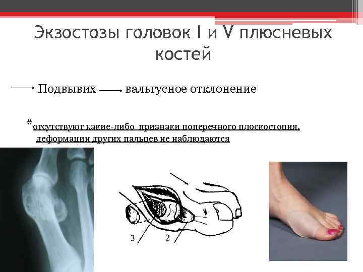 Exostoses of the heads of the I and V metatarsal bones Subluxation valgus deviation

The occurrence of hip deformities is based on various reasons. Part of the deformities comes from changes in the hip joint and femoral neck. Deformities in the area of the metaphysis and diaphysis of the thigh can be congenital, rachitic, inflammatory, can be associated with trauma and various tumors.
Rachitic hip deformities
A characteristic feature of the pathological process in the early period of rickets is the formation of osteoid tissue, which does not undergo timely ossification.
At the end of the disease, when the ossification process has not yet been fully restored, traction of the muscles, especially adductors, and premature load on the legs cause the curvature of the hips characteristic of rickets - the O-shaped thigh (femur varum). Bilateral hip deformity is more common.
Symptoms. Usually the deformity captures the entire thigh and lower leg.
Due to the arcuate deformity of the thigh and changes in the epiphyseal cartilage, the length of the limbs is reduced, there is a disproportion between the length of the trunk and limbs. The physiological axis of the femur is disturbed, and due to improper loading near the ankle joint, secondary deformity of the foot often occurs.
Prevention and treatment. In the period of fresh rickets, with a tendency to deformity of the limbs, it is necessary to fix them with a plaster splint and not allow the load on them until the bone structure is completely restored, which is checked radiologically. Temporarily give unloading orthopedic apparatus. At the same time, vitamin therapy and ultraviolet irradiation of the patient are carried out.
Treatment of the developed deformity of the hip consists of osteotomy, correction of its axis or lengthening.
The osteotomy is done under local anesthesia. The wide fascia, the external broad muscle, the periosteum are dissected with an external incision, the bleeding is carefully stopped. At the height of the greatest deformation of the thigh, an oblique osteotomy is made, skeletal traction is performed or plaster is applied for 2 months, then applied, therapeutic gymnastics, careful load in splint.
With a noticeable shortening of the limb due to hip deformity, it is possible to lengthen the entire limb in two ways: on the thigh or by surgery on the bones of the lower leg. To lengthen the femur, the method of segmental osteotomy according to N. A. Bogoraz is used with the introduction of a font into the medullary canal or Z-shaped osteotomy followed by skeletal traction.
Z-shaped osteotomy is performed as follows. After a Z-shaped dissection of the periosteum, the diaphysis is drilled with a narrow drill in the anteroposterior direction in 3-4 places, and make sure that the drill passes through the back wall.
Then, with a narrow sharp chisel, the femur is split along the length. The channels drilled before this make it possible to produce an osteotomy without any difficulty and of such a size as is required to eliminate the shortening of the femur.
After a Z-shaped osteotomy, some people drive an autograft into the bone marrow canal, which does not interfere with the stretching of the fragments, prevents their displacement and guarantees consolidation.
Then apply skin traction with a sticky patch, cleol, or zinc-gelatin paste with lateral pulls for 2 weeks to prevent lateral curvature.
The following complications are possible with limb lengthening:
- temporary muscle weakness from lengthening;
- fracture at the site of an earlier osteotomy;
- slow consolidation;
- vicious union;
- limited mobility in the knee after prolonged fixation.
Patients should be kept lying down for a long time, but with active movements in the joints and with emphasis on the legs. With proper postoperative management of the patient, complications can be avoided.
Limb lengthening can also be achieved by osteotomy of the tibia bones.
Recently, various screw devices, in particular the Gudushauri device, have been used to lengthen the femur and lower leg with good results.
Traumatic hip deformities
There are traumatic deformities of the upper third of the thigh, the area of the diaphysis and the distal end.
Symptoms. Hip deformity in the upper third occurs after damage to the epiphysis (epiphyseolysis), fracture of the neck (coxa vara traumatica) or the meta-diaphyseal part of the femur. In the latter case, an angular curvature of the femur develops with its shortening. In diaphyseal deformity of the femur, displacement of fragments along the length and width, violation of the physiological axis of the femur and shortening of the limb are the most important symptoms. Displacement of the distal fragment along the periphery and recurvation of the femur, outwardly hardly noticeable, significantly upset the function of the limb.
Treatment. In the indicated cases, the deformity is surgically eliminated by osteotomy and lengthening of the femur.
Hip deformities of inflammatory origin
Inflammatory processes that occur in the proximal or distal epiphysis of the femur in childhood lead to shortening of the limb and to a change in its shape and function.
The most significant deformations occur after the tuberculous process in the head and neck or in the distal epiphysis. Shortening in such cases sometimes reaches 8-10 cm or more. The shape and axis of the femur also change.
Similar hip deformities and shortening also develop after septic (metastatic) osteomyelitis of the femur that occurred in early childhood after umbilical sepsis.
Symptoms. The main symptoms are hip shortening and lameness. With a more thorough study, it is possible to detect abnormal development of the medial or lateral part of the distal epiphysis of the femur, a violation of its growth, sometimes premature synostosis and, as a result, the development of genu varum or genu valgum.
On the radiograph, it is possible to establish a violation of the structure of the meta-epiphyseal section and synostosis.
Treatment. Treatment of a shortened femur can be conservative or surgical. The use of orthopedic appliances or orthopedic shoes is indicated in children. Surgical lengthening of the femur is done with a shortening of more than 4 cm.
The article was prepared and edited by: surgeon