Cartilage cells. Skeletal (cartilaginous and bone) tissues
 The location of cartilage in the body n Cartilage tissue performs a shaping function in the fetus and a support in the adult body. Cartilaginous tissue can be found: n in the area of the joints (covering the articular surface with a relatively narrow layer), n in the metaphyses (i.e., between the epiphysis and diaphysis) of tubular bones, n in the intervertebral discs, in the anterior sections of the ribs, in the wall of the respiratory organs (larynx , trachea, bronchi), etc.
 Development n Like all other tissues of the internal environment of the body, skeletal tissues develop n from mesenchyme (the cells of which, in turn, are evicted from somites and splanchnotomes
 Features n The special nature of the intercellular substance gives two important properties: n elasticity and n strength. n of the intercellular substance of these tissues. n In many cases, cartilage is covered with perichondrium, a fibrous connective tissue that is involved in the growth and nutrition of cartilage.
 An important feature of cartilage tissue is the absence of blood vessels. Therefore, nutrients enter the cartilage - by diffusion from the vessels of the perichondrium. In some cases, there is no perichondrium - for example, in articular cartilage, since their surface should be smooth. Here, nutrition is carried out from the side of the synovial fluid and from the side of the underlying bone.
 Cellular composition n Chondroblasts are young cells, located in the deep layers of the perichondrium one by one and located closer to the surface of the cartilage n - small flattened cells capable of - proliferation and - synthesis of components of the intercellular substance of the cartilage. n granular EPS, Golgi complex, mitochondria are well expressed in them n Chondroblasts, releasing the components of the intercellular substance, "immure" themselves in it and turn into chondrocytes.
 Functions n The main function of chondroblasts is the production of the organic part of the intercellular substance: collagen and elastin proteins, glycosaminoglycans (GAGs) and proteoglycans (PGs). n Chondroblasts provide appositional (superficial) cartilage growth from the side of the perichondrium.
 Chondrocytes n a) Chondrocytes are the main type of cartilage cells. n - lie in special cavities of the intercellular substance (lacunae) and n - can divide by mitosis, while the daughter cells do not diverge, they remain together - isogenic groups (of 2-6 cells) are formed, originating from one cell. n b) They are n-larger (compared to chondroblasts) in size and oval in shape. n Well developed granular ER and Golgi complex
 Functions n Chondrocytes that have stopped dividing actively synthesize components of the intercellular substance. n Due to the activity of chondrocytes, an increase in the mass of cartilage from the inside occurs - interstitial growth.
 Chondroclasts n In the cartilaginous tissue, in addition to the cells forming the intercellular substance, there are also their antagonists - the destroyers of the intercellular substance - these are chondroclasts (can be attributed to the macrophage system): rather large cells, there are many lysosomes and mitochondria in the cytoplasm. Function - the destruction of damaged or worn sections of cartilage.
 Intercellular substance n The intercellular substance of cartilage tissue contains fibers and ground substance. n many fibrous structures: n-collagen fibers, n and in the elastic cartilage - elastic fibers.
 n The intercellular substance is highly hydrophilic, the water content reaches 75% of the mass of the cartilage, which leads to a high density and turgor of the cartilage. Cartilaginous tissues in the deep layers do not have blood vessels,
 n The main amorphous substance contains: n-water (70-80%), -mineral substances (4-7%), -organic component (10-15%), represented by n-proteoglycans and -glycoproteins.
 Proteoglycans n The proteoglycan aggregate contains 4 components. n At the heart of the aggregate is a long thread of hyaluronic acid (1). n With the help of globular binding proteins (2), n linear (fibrillar) peptide chains of the so-called. core (core) protein (3). n In turn, oligosaccharide branches (4) depart from the latter.
 These complexes n are highly hydrophilic; therefore, they bind a large amount of water and n provide high elasticity of the cartilage. n At the same time, they retain permeability to low molecular weight metabolites.
 n The perichondrium is a layer of connective tissue that covers the surface of cartilage. In the perichondrium, an external fibrous is isolated (from a dense, unformed CT with large quantity blood vessels) and the inner cell layer containing a large number of semi-stem cells.
 Hyaline cartilage n Outwardly, this tissue has a bluish-white color and looks like glass (Greek hyalos - glass). Hyaline cartilage - covers all articular surfaces of bones, is contained in the sternal ends of the ribs, in the airways.
 Distinctive features n 1. The intercellular substance of hyaline cartilage in preparations stained with hematoxylin-eosin seems to be homogeneous, not containing fibers. n 2. around the isogenic groups there is a clearly defined basophilic zone - the so-called territorial matrix. This is due to the fact that chondrocytes secrete a large amount of GAG with an acidic reaction, so this area is stained with basic colors, i.e., basophilic. Weakly oxyphilic areas between the territorial matrices are called the interterritorial matrix. n
 n A large number of proteoglycan aggregates. n Glycosaminoglycans. High elasticity depends on the content of GAGs n Chondroitin sulfates (chondroitin-6-sulfate, chondroitin-4-sulfate) n Keratan sulfates fibers). n Collagen IX, VI and X n Chondronectin protein
 Cellular composition n a) Immediately below the perichondrium are n young chondrocytes (3) - n are somewhat larger in size and more oval in shape. n b) Deeper are n mature chondrocytes n large oval cells with light cytoplasm, n forming isogenic groups (4) of 2-6 cells.
 n 1) Articular surfaces of bones. n 2) Airways. n 3) The junction of the ribs with the sternum.
 Elastic cartilage n In the auricle, epiglottis, cartilages of the larynx. In the intercellular substance, in addition to collagen fibers, there are a large number of randomly located elastic fibers, which gives elasticity to the cartilage. Elastic cartilage contains less lipids, chondroitin sulfates and glycogen.
 n b) in the thickness of the cartilaginous plate - isogenic groups of chondrocytes, n large, oval and n have a light cytoplasm. n Groups of chondrocytes usually have n-type chains (from 2, rarely more cells), oriented perpendicular to the surface.
 Age-related changes n Due to the relatively low content of collagen fibrils and the absence of collagen X, there is no deposition of calcium salts (calcification) in the elastic cartilage n in case of malnutrition.
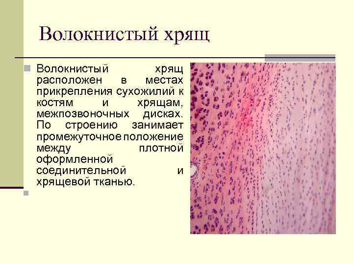 Fibrous cartilage n Fibrous cartilage is located at the points of attachment of tendons to bones and cartilage, intervertebral discs. In structure, it occupies an intermediate position between dense, formed connective and cartilage tissue. n
 n In the intercellular substance, there are much more collagen fibers arranged oriented - they form thick bundles that are clearly visible under a microscope. Chondrocytes often lie singly along the fibers without forming isogenic groups. They have an elongated shape, a rod-shaped nucleus and a narrow rim of the cytoplasm.
 n At the periphery, the fibrous cartilage gradually passes n into a dense, formed connective collagen fibers, which acquire orientation and go from one vertebra to another. tissue, oblique n b) In the central part of the disk, the fibrocartilage passes into the nucleus pulposus, which contains hyaline cartilage, type II collagen (in the form of fibrils)
 Cartilage regeneration n Hyaline - insignificant. The perichondrium is mainly involved n Elastic - less prone to degeneration and does not calcify n Fibrous - poor regeneration, capable of calcification

 Composition n Bone tissues consist of cells and intercellular substance. n Differenton of bone tissue includes n 1. stem and semi-stem (osteogenic) cells, n osteoblasts, n osteocytes n 2. osteoclasts.
 Osteoblasts n Osteoblasts are the most functionally active cellular elements of differon during osteohistogenesis. In an adult organism, the source of cells that support the population of osteoblasts are cells of the dispersed cambium in the osteogenic layer of the periosteum. Osteoblasts have a cubic or prismatic shape. The nucleus is located eccentrically. Osteoblasts are typical actively synthesizing and secreting cells; secretion is carried out by the entire surface of the cell. The cell has a well-developed granular endoplasmic reticulum that fills almost the entire cytoplasm, many free ribosomes and polysomes,
 Functions n secrete type I collagen, alkaline phosphatase, osteocalcin, osteopontin, transforming growth factors, osteonectin, collagenase, etc. n Highly differentiated osteoblasts are characterized by a gradual decrease in the activity of alkaline phosphatase, osteocalcin, osteopontin and the absence of proliferative activity.
 n Role in the mineralization of the organic basis of the bone matrix. The process of mineralization of the bone matrix begins with the deposition of amorphous calcium phosphate. Calcium cations enter the extracellular matrix from the bloodstream, where they are in a protein-bound state. n In the presence of alkaline phosphatase synthesized by osteoblasts, glycerophosphates in the intercellular substance are cleaved to form a phosphate anion. An excess of the latter leads to a local increase in Ca and P to a level at which calcium phosphate precipitates. The overwhelming fraction of the bone mineral is in the form of hydroxyapatite crystals. Crystals form on the collagen fibers of the bone matrix. The latter have structural features that contribute to this process. The fact is that the molecules of the precursor of collagen - tropocollagen are packed in a fiber in such a way that a gap remains between the end of one and the beginning of the other, called the zone of holes. It is in this zone that the bone mineral is initially deposited. Subsequently, the crystals begin to grow in both directions, and the process covers the entire fiber
 n A significant role in the mineralization of the synthesized organic bone matrix belongs to matrix vesicles. Such vesicles are derivatives of the Golgi complex of osteoblasts, have a membrane structure and contain various enzymes necessary for mineralization reactions or their inhibition, as well as amorphous calcium phosphates. Matrix vesicles exit the cells into the extracellular space and release the products contained in them. The latter initiate mineralization processes.
 Osteocytes n In terms of quantitative composition, the most numerous cells of bone tissue. These are process cells that lie in bone cavities - lacunae. The cell diameter reaches up to 50 microns. The cytoplasm is weakly basophilic. Organelles are poorly developed (granular EPS, PC and mitochondria). They don't share. n Function: take part in the physiological regeneration of bone tissue, produce the organic part of the intercellular substance. The thyroid hormone calcitonin has a stimulating effect on osteoblasts and osteocytes - the synthesis of the organic part of the intercellular substance increases and the deposition of calcium increases, while the concentration of calcium in the blood decreases.


 Osteoclasts n n n Specialized macrophages. Their diameter reaches up to 100 microns. Different compartments of osteoclasts are specialized for specific functions. the basal zone, in it, as part of numerous (5 - 20) nuclei, the genetic apparatus of the cell is concentrated. light area in direct contact with the bone matrix. Thanks to it, the osteoclast adheres tightly to the bone along the entire perimeter, creating an isolated space between itself and the surface of the mineralized matrix. Adhesion of the osteoclast is provided by a number of receptors to the components of the matrix, the main of which are receptors for vitronectin. The selective permeability of this barrier makes it possible to create a specific microenvironment in the cell adhesion zone. the vesicular zone contains lysosomes. Enzymes, acidic substances are transported through the membrane of the corrugated border, carbonic acid H 2 CO 3 is formed; carbonic acid dissolves calcium salts, dissolved calcium is washed into the blood. carrying out demineralization and disorganization of the bone matrix, which leads to the formation of a resorption (erosive) Hausship lacunae.
 Osteoclasts n osteoclasts have many nuclei and a large amount of cytoplasm; the zone of cytoplasm adjacent to the bone surface is called the corrugated border, there are many cytoplasmic outgrowths and lysosomes functions - the destruction of fibers and amorphous bone substance
 n Thick collagen fibers, devoid of cementing substance, create a "brush border" appearance. Lysosomal enzymes proteolyze collagen and other matrix proteins. Proteolysis products are removed from osteoclastic lacunae by transcellular transport. In general, the process of reducing the river. H in the lacuna is carried out by two mechanisms: by exocytosis of the acidic contents of the vacuoles into the lacuna and due to the action of proton pumps - H + -ATPases localized in the membrane of the corrugated border. The source for hydrogen ions is water and carbon dioxide, which are the result of mitochondrial oxidation reactions.
 Intercellular substance n 1. The inorganic part of the matrix It contains calcium (35%) and phosphorus (50%) (calcium phosphate and carbonate salts) mainly in the form of hydroxyapatite crystals (Ca 10 (PO 4) 6 (OH) 2 ) (3 Ca (OH) 2), n and a little - in the amorphous state, a small amount of magnesium phosphate - make up 70% of the intercellular substance. In plasma, inorganic phosphorus is contained in the form of anions HPO 4 -2 and H 2 PO 4 -2. n n The ratio of the organic and inorganic parts of the intercellular substance depends on age: in children, the organic part is slightly more than 30%, and the inorganic part is less than 70%, so their bones are less strong, but more flexible (not brittle); in old age, on the contrary, the proportion the inorganic part increases and the organic part decreases, so the bones become harder but more brittle - blood vessels are present:
 The organic part of the bone matrix The organic part of the intercellular substance is represented by n collagen (collagen types I, X, V), very few glycosaminoglycans and proteoglycans. n - glycoproteins (alkaline phosphatase, osteonectin); n - proteoglycans (acid polysaccharides and glycosaminoglycans - chondroitin-4 - and chondroitin-6 sulfates, dermatan sulfate and keratan sulfate.); n - growth factors (fibroblast growth factor, transforming growth factors, bone morphogenetic proteins) - cytokines secreted by bone tissue and blood cells, which carry out local regulation of osteogenesis.
 proteins that carry out cell adhesion n Osteonectin is a glycoprotein of bone and dentin, has a high affinity for type I collagen and hydroxyapatite, contains Ca-binding domains. It maintains the concentration of Ca and P in the presence of collagen. It is assumed that the protein is involved in the interaction of the cell and the matrix. n Osteopontin is the main component of the protein composition of the matrix, in particular interfaces, where it accumulates in the form of a dense cover called cementation lines (lamina limitans). Due to its physicochemical properties, it regulates the calcification of the matrix, specifically participates in the adhesion of cells to the matrix or matrix to the matrix. The production of osteopontin is one of the earliest manifestations of osteoblast activity. n Osteocalcin (OC) - a small protein (5800 Da, 49 amino acids) in the mineralized bone matrix, is involved in the process of calcification,
 Classification n There are tubular, flat and mixed bones. The diaphyses of tubular bones and the cortical plates of flat and mixed bones are built from lamellar bone tissue covered with periosteum or periosteum. In the periosteum, it is customary to distinguish two layers: the outer one is fibrous, consisting mainly of fibrous connective tissue; internal, adjacent to the surface of the bone - osteogenic, or cambial.
 Types of bone tissue Coarse-fibrous (reticulofibrous) lamellar (fine-fibrous) The main feature Collagen fibers form a) Bone substance is thick bundles running in different (organized into plates). directions. b) Moreover, within the same plate, the fibers have the same direction, and within neighboring plates, they are different. Localization 1. Flat bones of the embryo. 2. Tubercles of bones; sites of overgrown cranial sutures. Almost all bones of an adult: flat (scapula, pelvic bones, skull bones), spongy (ribs, sternum, vertebrae) and tubular.

 Lamellar bone tissue can have a spongy and compact organization. Cancellous bone substance Compact bone substance Localization Spongy substance consists of: the epiphyses of tubular bones, the inner layer (adjacent to the medullary canal) of the diaphysis of tubular bones, spongy bones, the inner part of flat bones. Most of the diaphyses of tubular bones and the surface layer of flat bones have a compact structure. Distinctive feature The spongy substance is built from avascular bone beams (beams), between which there are gaps - bone cells. There are practically no gaps in the compact bone substance: due to the growth of bone tissue deep into the cells, only narrow spaces for blood vessels remain - the so-called. central canals of osteons Bone marrow The cells of the spongy substance contain vessels that feed the bone, and red bone marrow is a hematopoietic organ. The medullary cavity of the diaphysis of tubular bones in adults contains yellow bone marrow - adipose tissue.
 Structure They consist of bone plates a) In this case, the plates of the spongy substance are usually oriented along the direction of the bone beams, and not around the vessels, as in osteons of a compact substance. b) osteons can occur in sufficiently thick beams. The unit of structure is the bone plates. They consist of bone plates. In a compact substance, there are plates of 3 types: general (general) - surround the entire bone, osteon - lie in concentric layers around the vessel, forming the so-called. osteons; intercalary - located between osteons. osteons.
 The structure of the osteon, the main structural unit of the bone In the center of each osteon is a blood vessel (1), around the latter there are several concentric layers of bone plates (2), called osteons. Osteons are delimited by a resorption (spinal) line (3). Intercalated bone plates (4) lie between the osteons, which are the remnants of previous generations of osteons. bone plates include cells (osteocytes), collagen fibers and a ground substance rich in mineral compounds. the fibers in the intercellular substance are indistinguishable, and the intercellular substance itself has a solid consistency.

 BONE DEVELOPMENT FROM MESENCHYME (direct osteohistogenesis). From the mesenchyme, an immature (coarse-fibred) bone is formed, which is subsequently replaced by a lamellar bone. There are 4 stages in development: n 1. formation of an osteogenic island - in the area of bone formation, mesenchymal cells turn into osteoblasts n
 2. formation of intercellular substance n osteoblasts begin to form the intercellular substance of the bone, while some of the osteoblasts are inside the intercellular substance, these osteoblasts turn into osteocytes; the other part of the osteoblasts is on the surface of the intercellular substance,
 3. Calcification of the intercellular substance of the bone The intercellular substance is impregnated with calcium salts. n a) At the third stage, so-called. matrix vesicles similar to lysosomes. They accumulate calcium and (due to alkaline phosphatase) inorganic phosphate. n b) When the bubbles burst, mineralization of the intercellular substance occurs, i.e., the deposition of hydroxyapatite crystals on the fibers and in the amorphous substance. As a result, bone trabeculae (beams) are formed - mineralized tissue areas containing all 3 types of bone cells - n n n from the surface - osteoblasts and osteoclasts, and in depth - osteocytes.
 4. Formation of osteons n Subsequently, in the inner part of the flat bone n, the primary spongy tissue is replaced by a secondary one, n which is already built from bone plates oriented along the beams.
 The development of lamellar bone tissue is closely related to 1. the process of destruction of individual sections of the bone and the ingrowth of blood vessels into the thickness of the reticulofibrous bone. Osteoclasts are involved in this process both during embryonic osteogenesis and after birth. 2. vessels growing to the trabeculae. In particular, around the vessels, the bone substance is formed in the form of concentric bone plates that make up the primary osteons.
 DEVELOPMENT OF THE BONE IN THE SITE OF THE CARTILAGE (indirect osteogenesis) n in place of the cartilage, a mature (lamellar) bone is immediately formed n 4 stages are distinguished in development: n 1. formation of cartilage - in place of the future bone, hyaline cartilage is formed
 2. perichondral ossification takes place only in the area of the diaphysis in the area of the diaphysis, the perichondrium turns into the periosteum, in which osteogenic cells appear, then osteoblasts, due to the osteogenic cells of the periosteum, on the surface of the cartilage, bone formation begins in the form of common plates that have a circular course, like the annual rings of a tree
 3. endochondral ossification n Occurs both in the area of the diaphysis and in the area of the epiphysis; blood vessels grow inside the cartilage, where there are osteogenic cells - osteoblasts, due to which bone is formed around the vessels in the form of osteons, and osteoclasts. n simultaneously with the formation of bone, the destruction of cartilage occurs
 zone of vesicular cartilage (4). At the border of the still preserved cartilage, the cartilage cells are in a swollen, vacuolated state, i.e., they have a bubble-shaped zone of columnar cartilage (5). In the adjacent region of the epiphysis, cartilage continues to grow and the proliferating cells line up in columns along the long axis of the bone.
 n a) Subsequently, ossification of the epiphysis itself (with the exception of the articular surface) will occur - by the endochondral way. n b) That is, mineralization will also occur here, n vessels will sprout here, the substance of the cartilage will collapse and first coarse fibrous, n and then lamellar bone tissue will form.
 n 4. restructuring and growth of the bone - the old parts of the bone are gradually destroyed and new ones are formed in their place; due to the periosteum, common bone plates are formed, due to the osteogenic cells located in the adventitia of the vessels of the bone, osteons are formed. Between the diaphysis and the epiphysis, a layer of cartilaginous tissue is preserved, due to which the growth of the bone in length continues until the end of the period of growth of the body in length, i.e. up to 20-21 years.
 Bone growth Sources of growth Until the age of 20, tubular bones grow: in width - by appositional growth from the side of the perichondrium, in length - due to the activity of the metaepiphyseal cartilaginous plate. Metaepiphyseal cartilage a) Metaepiphyseal plate - a part of the epiphysis adjacent to the diaphysis and retaining (unlike the rest of the epiphysis) the cartilaginous structure. b) It has 3 zones (in the direction from the epiphysis to the diaphysis): the border zone - contains oval chondrocytes, the zone of columnar cells - it is this that ensures the growth of cartilage in length due to the multiplication of chondrocytes, the zone of vesicular cartilage - borders on the diaphysis and undergoes ossification . c) Thus, 2 processes simultaneously occur: cartilage growth (in the columnar zone) and its replacement with bone (in the vesicular zone).
 Regeneration n Regeneration and growth of the bone in thickness is carried out due to the periosteum and endosteum. All tubular bones, as well as most flat bones, are histologically fine-fibered bone.
 n In the bone tissue, two oppositely directed processes constantly occur - resorption and neoplasm. The ratio of these processes depends on several factors, including age. Restructuring of bone tissue is carried out in accordance with the loads acting on the bone. n The process of bone tissue remodeling occurs in several phases, in each of which certain cells play the leading role. Initially, the area of bone tissue to be resorbed is "marked" by osteocytes using specific cytokines (activation). The protective layer on the bone matrix is destroyed. Precursors of osteoclasts migrate to the bare surface of the bone, merge into a multinuclear structure - a symplast - a mature osteoclast. At the next stage, the osteoclast demineralizes the bone matrix (resorption), gives way to macrophages, which complete the destruction of the organic matrix of the bone intercellular substance and prepare the surface for osteoblast adhesion (reversion). At the last stage, precursors arrive in the destruction zone, differentiating into osteoblasts, they synthesize and mineralize the matrix in accordance with the new conditions of static and dynamic load on the bone (formation).
Cartilage tissue includes 3 types of cartilage (hyaline, elastic and fibrous), differing from each other mainly in the structure of the intercellular substance. There are no blood vessels in the cartilage tissue, therefore its trophism is carried out diffusely due to the vessels of the perichondrium or synovial fluid.
Cells: chondroblasts, chondrocytes and chondroclasts.
Chondroblasts- poorly differentiated cells of cartilage tissue, in embryogenesis are formed from undifferentiated mesenchymal cells; have an oval shape, sometimes with pointed ends. In their basophilically stained cytoplasm, HES is well developed, which is associated with the synthesis of proteins in the intercellular substance of the cartilage. Under certain circumstances, they are able to produce enzymes that destroy the intercellular substance - collagenase, elastase, hyaluronidase. They are localized in the cartilage growth zones (in the inner layer of the perichondrium). As chondroblasts age, the amount of granular endoplasmic reticulum decreases and they turn into chondrocytes.
Chondrocytes- differentiated cartilage cells, the shape of which is already becoming rounded or angular. The synthesis of the intercellular substance of the cartilage in them proceeds at a lower level than in chondroblasts. They are located in the thickness of the intercellular substance in special cavities - lacunae. Sometimes in one gap there are several chondrocytes, which were formed as a result of the division of one cell that has not yet lost the ability to mitosis. Therefore, such groups of cells are called isogenic.
Chondroclasts- a type of polynuclear macrophages that are involved in the destruction of cartilage.
intercellular substance represented by an amorphous component and fibers. Hyaline and fibrous cartilage contain only collagen (chondrin) fibers, while elastic cartilage contains predominantly elastic and, to a lesser extent, collagen. The amorphous component is represented by proteoglycans and glycosaminoglycans.
Localization:
Hyaline cartilage - in the trachea and bronchi, articular surfaces, in the larynx, connections of the ribs with the sternum;
Elastic - in the auricles, carob-shaped and sphenoid cartilages of the larynx, cartilages of the nose;
Fibrous cartilage - in places where tendons and ligaments pass into hyaline cartilage, in intervertebral discs, semi-movable joints, symphyses. So, for example, in the intervertebral disc there is a nucleus pulposus inside, consisting of glycosaminoglycans and proteoglycans and cartilage cells localized in them, and outside there is a fibrous ring, which contains mainly fibers that have a circular course.
perichondrium consists of 2 layers. Its outer layer is formed by a dense fibrous unformed connective tissue, and the inner (chondrogenic) layer is formed by loose fibrous connective tissue, in which there are many chondroblasts and blood vessels. Due to the inner layer, trophism and regeneration of cartilage tissue is carried out.
cartilage growth is carried out in two ways: due to the chondrogenic layer of the perichondrium (appositional growth) and due to the reproduction of cells located in cavities inside the cartilage, which have not yet lost the ability to divide (internal, or interstitial growth).
Histogenesis of cartilage tissue is carried out from mesenchymocytes, which are evicted from sclerotomes, which form chondrogenic islets. The differentiation of mesenchymocytes into chondrogenic cells and chondroblasts is accompanied by the synthesis of an intercellular substance that fills the gaps between cells, separating them from each other. The cells separated in this way are still able to divide for some time and turn into chondrocytes, which are located in isogenic groups in one gap.
70. Cartilaginous tissues. Classification, development, structure, histochemical characteristics and function. Cartilage growth, regeneration and age-related changes.
cartilaginous and bone tissue develop from the sclerotomic mesenchyme, belong to the tissues of the internal environment and, like all other tissues of the internal environment, consist of cells and intercellular substance. The intercellular substance here is dense, so these tissues perform a support-mechanical function.
cartilage tissue(textuscartilagineus). They are classified into hyaline, elastic and fibrous. The classification is based on the features of the organization of the intercellular substance. The composition of cartilage tissue includes 80% water, 10-15% organic matter and 5-7% inorganic matter.
Cartilage development, or chondrogenesis, consists of 3 stages: 1) the formation of chondrogenic islets; 2) formation of primary cartilaginous tissue; 3) differentiation of cartilaginous tissue.
During 1st stage mesenchymal cells combine into chondrogenic islets, the cells of which multiply, differentiate into chondroblasts. The formed chondroblasts contain granular EPS, the Golgi complex, and mitochondria. The chondroblasts then differentiate into chondrocytes.
During 2nd stage in chondrocytes, granular EPS, the Golgi complex, and mitochondria are well developed. Chondrocytes actively synthesize fibrillar protein (collagen type II), from which an intercellular substance is formed that stains oxyphilically.
On the onset 3rd stage in chondrocytes, granular ER develops more intensively, on which both fibrillar proteins and chondroitin sulfates (chondroitin sulfuric acid) are produced, which are stained with basic dyes. Therefore, the main intercellular substance of the cartilaginous tissue around these chondrocytes is stained basophilically.
A perichondrium is formed around the cartilaginous rudiment from mesenchymal cells, consisting of 2 layers: 1) outer, denser, or fibrous, and 2) inner, looser, or chondrogenic, which contains prechondroblasts and chondroblasts.
appositional growth of cartilage or growth by superposition, is characterized by the fact that chondroblasts are released from the perichondrium, which are superimposed on the main substance of the cartilage, differentiate into chondrocytes and begin to produce the intercellular substance of the cartilage tissue.
Interstitial growth cartilage tissue is carried out due to chondrocytes located inside the cartilage, which, firstly, divide by mitosis and, secondly, produce an intercellular substance, due to which the volume of cartilage tissue increases.
Cartilage cells(chondrocytus). The chondrocyte differon is composed of: stem cell, half-stem cell (prechondroblast), chondroblast, chondrocyte.
Chondroblasts (chondroblastus) are located in the inner layer of the perichondrium, have organelles of general importance: granular ER, Golgi complex, mitochondria. Functions of chondroblasts:
1) secrete intercellular substance (fibrillar proteins);
2) in the process of differentiation they turn into chondrocytes;
3) have the ability to mitotic division.
Chondrocytes located in cartilaginous lacunae. In the lacuna, at first, there is 1 chondrocyte, then, in the process of its mitotic division, 2, 4, 6, etc. cells are formed. All of them are located in the same lacuna and form an isogenic group of chondrocytes.
Chondrocytes of the isogenic group are divided into 3 types: I, II, III.
Type I chondrocytes have the ability to mitotic division, contain the Golgi complex, mitochondria, granular ER and free ribosomes, have a large nucleus and a small amount of cytoplasm (large nuclear-cytoplasmic ratio). These chondrocytes are located in young cartilage.
Type II chondrocytes located in mature cartilage, their nuclear-cytoplasmic ratio decreases somewhat, as the volume of the cytoplasm increases; they lose the ability to mitosis. In their cytoplasm, granular ER is well developed; they secrete proteins and glycosaminoglycans (chondroitin sulfates), so the main intercellular substance around them stains basophilically.
Chondrocytes III type are located in the old cartilage, lose the ability to synthesize glycosaminoglycans and produce only proteins, so the intercellular substance around them stains oxyphilically. Therefore, a ring stained oxyphilically (proteins are isolated by type III chondrocytes) is visible around such an isogenic group, a basophilically stained ring is visible outside of this ring (glycosaminoglycans are secreted by type II chondrocytes) and the outer ring itself is again stained oxyphilically (proteins are isolated at a time when in cartilage contained only young type I chondrocytes). Thus, these 3 differently colored rings around isogenic groups characterize the process of formation and function of chondrocytes of 3 types.
Intercellular substance of cartilaginous tissue. Contains organic substances (mainly type II collagen), glycosaminoglycans, proteoglycans and non-collagen type proteins. The more proteoglycans, the more hydrophilic the intercellular substance, the more elastic and more permeable it is. Gases, water molecules, salt ions and micromolecules diffusely penetrate through the main substance from the side of the perichondrium. However, macromolecules do not penetrate. Macromolecules have antigenic properties, but since they do not penetrate cartilage, cartilage transplanted from one person to another takes root well (no immune rejection reaction occurs).
In the ground substance of cartilage there are collagen fibers, consisting of type II collagen. The orientation of these fibers depends on the lines of force, and the direction of the latter depends on the mechanical effect on the cartilage. There are no blood and lymphatic vessels in the intercellular substance of the cartilage tissue, therefore, the nutrition of the cartilage tissue is carried out by diffuse intake of substances from the vessels of the perichondrium.
Age-related changes in cartilage. The greatest changes are observed in old age, when the number of chondroblasts in the perichondrium and the number of dividing cartilage cells decrease. In chondrocytes, the amount of granular EPS, the Golgi complex and mitochondria decreases, the ability of chondrocytes to synthesize glycosaminoglycans and proteoglycans is lost. A decrease in the amount of proteoglycans leads to a decrease in the hydrophilicity of the cartilage tissue, a weakening of the permeability of the cartilage and the supply of nutrients. This leads to calcification of the cartilage, the penetration of blood vessels into it and the formation of bone substance inside the cartilage.
Cartilaginous tissue (textus cartilaginus) forms articular cartilages, intervertebral discs, cartilages of the larynx, trachea, bronchi, external nose. Consists cartilage tissue from cartilage cells (chondroblasts and chondrocytes) and dense, elastic intercellular substance.
Cartilaginous tissue contains about 70-80% water, 10-15% organic matter, 4-7% salts. About 50-70% of the dry matter of cartilage tissue is collagen. The intercellular substance (matrix) produced by cartilage cells consists of complex compounds, which include proteoglycans. hyaluronic acid, glycosaminoglycan molecules. There are two types of cells in the cartilaginous tissue: chondroblasts (from the Greek chondros - cartilage) and chondrocytes.
Chondroblasts are young, capable of mitotic division, rounded or ovoid cells. They produce components of the intercellular substance of cartilage: proteoglycans, glycoproteins, collagen, elastin. The cytolemma of chondroblasts forms many microvilli. The cytoplasm is rich in RNA, a well-developed endoplasmic reticulum (granular and non-granular), the Golgi complex, mitochondria, lysosomes, and glycogen granules. The chondroblast nucleus, rich in active chromatin, has 1-2 nucleoli.
Chondrocytes are mature large cartilage cells. They are round, oval or polygonal, with processes, developed organelles. Chondrocytes are located in cavities - lacunae, surrounded by intercellular substance. If there is one cell in the gap, then such a gap is called primary. Most often, the cells are located in the form of isogenic groups (2-3 cells) occupying the cavity of the secondary lacuna. The walls of the lacunae consist of two layers: the outer one, formed by collagen fibers, and the inner one, consisting of aggregates of proteoglycans that come into contact with the glycocalyx of cartilage cells.
The structural and functional unit of cartilage is the chondron, formed by a cell or an isogenic group of cells, a pericellular matrix, and a lacuna capsule.
Cartilage tissue is nourished by diffusion of substances from the blood vessels of the perichondrium. Nutrients enter the articular cartilage tissue from the synovial fluid or from the vessels of the adjacent bone. Nerve fibers are also localized in the perichondrium, from where individual branches of amyopiatic nerve fibers can penetrate into the cartilaginous tissue.
In accordance with the structural features of the cartilage tissue, there are three types of cartilage: hyaline, fibrous and elastic cartilage.
hyaline cartilage, from which the cartilages of the respiratory tract, the thoracic ends of the ribs and the articular surfaces of the bones are formed in humans. In a light microscope, its main substance appears to be homogeneous. Cartilage cells or their isogenic groups are surrounded by an oxyphilic capsule. In differentiated areas of cartilage, a basophilic zone adjacent to the capsule and an oxyphilic zone located outside of it are distinguished; Together, these zones form a cellular territory, or chondrin ball. The complex of chondrocytes with a chondrin ball is usually mistaken for functional unit cartilaginous tissue - chondron. The ground substance between chondrons is called interterritorial spaces.
Elastic cartilage(synonym: mesh, elastic) differs from hyaline by the presence of branching networks of elastic fibers in the main substance. The cartilage of the auricle, epiglottis, vrisberg and santorin cartilages of the larynx are built from it.
fibrocartilage(a synonym for connective tissue) is located at the transition points of dense fibrous connective tissue into hyaline cartilage and differs from the latter by the presence of real collagen fibers in the ground substance.
7. Bone tissue - location, structure, functions
Bone tissue is a type of connective tissue and consists of cells and intercellular substance, which contains a large amount of mineral salts, mainly calcium phosphate. Minerals make up 70% of bone tissue, organic - 30%.
Functions of bone tissue:
1) support;
2) mechanical;
3) protective (mechanical protection);
4) participation in the mineral metabolism of the body (depot of calcium and phosphorus).
Bone cells - osteoblasts, osteocytes, osteoclasts. The main cells in the formed bone tissue are osteocytes. These are process-shaped cells with a large nucleus and weakly expressed cytoplasm (nuclear-type cells). The cell bodies are localized in the bone cavities (lacunae), and the processes are located in the bone tubules. Numerous bone tubules, anastomosing with each other, penetrate the bone tissue, communicating with the perivascular space, form the drainage system of the bone tissue. In this drainage system contains tissue fluid, through which the exchange of substances is ensured not only between cells and tissue fluid, but also in the intercellular substance.
Osteocytes are definitive forms of cells and do not divide. They are formed from osteoblasts.
osteoblasts found only in developing bone tissue. In the formed bone tissue, they are usually contained in an inactive form in the periosteum. In developing bone tissue, osteoblasts surround each bone plate along the periphery, tightly adhering to each other.
The shape of these cells can be cubic, prismatic and angular. The cytoplasm of osteoblasts contains a well-developed endoplasmic reticulum, the Golgi lamellar complex, many mitochondria, which indicates a high synthetic activity of these cells. Osteoblasts synthesize collagen and glycosaminoglycans, which are then released into the extracellular space. Due to these components, an organic matrix of bone tissue is formed.
These cells provide mineralization of the intercellular substance through the release of calcium salts. Gradually releasing the intercellular substance, they seem to be walled up and turn into osteocytes. At the same time, intracellular organelles are significantly reduced, synthetic and secretory activity is reduced, and the functional activity characteristic of osteocytes is preserved. Osteoblasts localized in the cambial layer of the periosteum are in an inactive state; synthetic and transport organelles are poorly developed in them. When these cells are irritated (in case of injuries, bone fractures, etc.), a granular ER and a lamellar complex rapidly develop in the cytoplasm, active synthesis and release of collagen and glycosaminoglycans, the formation of an organic matrix (bone callus), and then the formation of a definitive bone fabrics. In this way, due to the activity of osteoblasts of the periosteum, bones regenerate when they are damaged.
osteoclasts- bone-destroying cells are absent in the formed bone tissue, but are contained in the periosteum and in places of destruction and restructuring of bone tissue. Since local processes of bone tissue restructuring are continuously carried out in ontogeny, osteoclasts are also necessarily present in these places. In the process of embryonic osteohistogenesis, these cells play a very important role and are present in large numbers. Osteoclasts have a characteristic morphology: these cells are multinucleated (3-5 or more nuclei), have a rather large size (about 90 microns) and a characteristic shape - oval, but the part of the cell adjacent to the bone tissue has a flat shape. In the flat part, two zones can be distinguished: the central (corrugated part, containing numerous folds and processes), and the peripheral part (transparent) in close contact with the bone tissue. In the cytoplasm of the cell, under the nuclei, there are numerous lysosomes and vacuoles of various sizes.
The functional activity of the osteoclast is manifested as follows: in the central (corrugated) zone of the cell base, carbonic acid and proteolytic enzymes are released from the cytoplasm. The released carbonic acid causes demineralization of bone tissue, and proteolytic enzymes destroy the organic matrix of the intercellular substance. Fragments of collagen fibers are phagocytosed by osteoclasts and destroyed intracellularly. Through these mechanisms, resorption (destruction) of bone tissue occurs, and therefore osteoclasts are usually localized in the depressions of bone tissue. After the destruction of bone tissue due to the activity of osteoblasts, which are evicted from the connective tissue of the vessels, a new bone tissue is built.
intercellular substance bone tissue consists of the main (amorphous) substance and fibers, which contain calcium salts. The fibers consist of collagen and are folded into bundles, which can be arranged in parallel (orderly) or randomly, on the basis of which the histological classification of bone tissues is built. The main substance of bone tissue, as well as other types of connective tissues, consists of glycosamino- and proteoglycans.
The bone tissue contains less chondroitin sulfuric acids, but more citric and others, which form complexes with calcium salts. In the process of bone tissue development, an organic matrix is first formed - the main substance and collagen fibers, and then calcium salts are deposited in them. They form crystals - hydroxyapatites, which are deposited both in an amorphous substance and in fibers. Providing bone strength, calcium phosphate salts are also both a depot of calcium and phosphorus in the body. Thus, bone tissue takes part in the mineral metabolism of the body.
When studying bone tissue, one should also clearly separate the concepts of “bone tissue” and “bone”.
Bone is an organ whose main structural component is bone tissue.
Classification of bone tissue
Cartilage tissue is a type of connective tissue, consisting of cartilage cells (chondrocytes) and a large amount of dense intercellular substance. Acts as a support. Chondrocytes have a variety of shapes and lie singly or in groups within cartilage cavities. The intercellular substance contains chondrin fibers, similar in composition to collagen fibers, and the main substance, rich in chondromucoid.
Depending on the structure of the fibrous component of the intercellular substance, three types of cartilage are distinguished: hyaline (vitreous), elastic (mesh) and fibrous (connective tissue).
Cartilage pathology - see Chondritis, Chondrodystrophy.
Cartilaginous tissue (tela cartilaginea) is a type of connective tissue characterized by the presence of a dense intercellular substance. In the latter, the main amorphous substance is distinguished, which contains compounds of chondroitinsulfuric acid with proteins (chondromucoids) and chondrin fibers, similar in composition to collagen fibers. Fibrils of cartilaginous tissue belong to the type of primary fibers and have a thickness of 100-150 Å. Electron microscopy in the fibers of the cartilaginous tissue, in contrast to the actual collagen fibers, reveals only an indistinct alternation of light and dark areas without a clear periodicity. Cartilage cells (chondrocytes) are located in the cavities of the ground substance singly or in small groups (isogenic groups).
The free surface of the cartilage is covered with dense fibrous connective tissue - the perichondrium (perichondrium), in the inner layer of which there are poorly differentiated cells - chondroblasts. The cartilaginous tissue of the perichondrium that covers the articular surfaces of the bones does not have. The growth of cartilage tissue is carried out due to the reproduction of chondroblasts, which produce the ground substance and later turn into chondrocytes (appositional growth) and due to the development of a new ground substance around chondrocytes (interstitial, intussusceptive growth). During regeneration, the development of cartilage tissue can also occur by homogenizing the basic substance of the fibrous connective tissue and converting its fibroblasts into cartilage cells.
Cartilage tissue is nourished by diffusion of substances from the blood vessels of the perichondrium. Nutrients enter the articular cartilage tissue from the synovial fluid or from the vessels of the adjacent bone. Nerve fibers are also localized in the perichondrium, from where individual branches of amyopiatic nerve fibers can penetrate into the cartilaginous tissue.
In embryogenesis, cartilaginous tissue develops from mesenchyme (see), between the approaching elements of which layers of the main substance appear (Fig. 1). In such a skeletal rudiment, hyaline cartilage is first formed, temporarily representing all the main parts of the human skeleton. In the future, this cartilage can be replaced by bone tissue or differentiate into other types of cartilage tissue.
The following types of cartilage tissue are known.
hyaline cartilage(Fig. 2), from which the cartilages of the respiratory tract, the thoracic ends of the ribs and the articular surfaces of the bones are formed in humans. In a light microscope, its main substance appears to be homogeneous. Cartilage cells or their isogenic groups are surrounded by an oxyphilic capsule. In differentiated areas of cartilage, a basophilic zone adjacent to the capsule and an oxyphilic zone located outside of it are distinguished; Together, these zones form a cellular territory, or chondrin ball. A complex of chondrocytes with a chondrin ball is usually taken as a functional unit of cartilage tissue - a chondron. The ground substance between chondrons is called interterritorial spaces (Fig. 3).
Elastic cartilage(synonym: reticulate, elastic) differs from hyaline by the presence of branching networks of elastic fibers in the ground substance (Fig. 4). The cartilage of the auricle, epiglottis, vrisberg and santorin cartilages of the larynx are built from it.
fibrocartilage(a synonym for connective tissue) is located at the transition sites of dense fibrous connective tissue into hyaline cartilage and differs from the latter by the presence of real collagen fibers in the ground substance (Fig. 5).
Cartilage pathology - see Chondritis, Chondrodystrophy, Chondroma.
Rice. 1-5. The structure of cartilage.
Rice. 1. Cartilage histogenesis:
1 - mesenchymal syncytium;
2 - young cartilage cells;
3 - layers of the main substance.
Rice. 2. Hyaline cartilage (small magnification):
1 - perichondrium;
2 - cartilage cells;
3 - the main substance.
Rice. 3. Hyaline cartilage (large magnification):
1 - isogenic group of cells;
2 - cartilaginous capsule;
3 - basophilic zone of the chondrin ball;
4 - oxyphilic zone of the chondrin ball;
5 - interterritorial space.
Rice. 4. Elastic cartilage:
1 - elastic fibers.
Rice. 5. Fibrous cartilage.
The bone marrow filling the marrow cavities contains mainly fats (up to 98% in the dry residue of the yellow marrow) and less choline phosphatides, cholesterol, proteins and minerals. The composition of fats is dominated by palmitic, oleic, stearic acids.
In accordance with the characteristics of the chemical composition, the bone is used for the production of semi-finished products, jelly, brawn, bone fat, gelatin, glue, bone meal.
cartilage tissue. Cartilage tissue performs supporting and mechanical functions. It consists of a dense ground substance, in which round-shaped cells, collagen and elastin fibers are located (Fig. 5.14). Depending on the composition of the intercellular substance, hyaline, fibrous and elastic cartilages are distinguished. Hyaline cartilage covers the articular surfaces of the bones, costal cartilages and the trachea are built from it. Calcium salts are deposited in the intercellular substance of such cartilage with age. Hyaline cartilage is translucent, has a bluish tint. 
Fibrous cartilage is made up of ligaments between vertebrae, as well as tendons and ligaments where they attach to bones. Fibrous cartilage contains many collagen fibers and a small amount of amorphous matter. It has the appearance of a translucent mass.
Cream-colored elastic cartilage, in the intercellular substance of which elastin fibers predominate. Lime is never deposited in elastic cartilage.
cartilage tissue
It is part of the auricle, larynx.
Average chemical composition cartilage tissue includes: 40-70% water, 19-20% proteins, 3.5% fats, 2-10% minerals, about 1% glycogen.
Cartilage tissue is characterized by a high content of mucoprotein - chondromucoid and mucogyulisaccharide - chondroitinsulfuric acid in the main intercellular substance. An important property of this acid is its ability to form salt-like compounds with various proteins: collagen, albumin, etc. This, apparently, explains the "cementing" role of mucopolysaccharides in cartilage tissue.
Cartilage tissue is used for food purposes, and gelatin and glue are also produced from it. However, the quality of gelatin and glue is often not high enough, since mucopolysaccharides and glucoproteins pass into solution from the tissue along with gelatin, reducing the viscosity and strength of the jelly.
Cartilage tissues are a type of supporting tissues characterized by the strength and elasticity of the matrix. This is due to their position in the body: in the area of the joints, in the intervertebral discs, in the wall of the respiratory tract (larynx, trachea, bronchi).
cartilaginous
○ Hyaline
○ Elastic
○ Fibrous
However, the general plan of their structure is similar.
1. The presence of cells (chondrocytes and chondroblasts).
2. Formation of isogenic groups of cells.
3. The presence of a large amount of intercellular substance (amorphous, fibers), which provides strength and elasticity - that is, the ability to reversibly deform.
4. Absence of blood vessels - nutrients diffuse from the perichondrium due to the high water content (up to 70-80%) in the matrix.
5. Characterized by a relatively low level of metabolism.
cartilage tissue
They have the ability to grow continuously.
In the process of development of cartilage tissue, a differon of cartilage cells is formed from the mesenchyme. It includes:
1. Stem cells - are characterized by a rounded shape, a high value of nuclear-cytoplasmic ratios, a diffuse arrangement of chromatin and a small nucleolus. Cytoplasmic organelles are poorly developed.
2. Semi-stem cells (prechondroblasts) - the number of free ribsomes increases in them, rEPS appears, the cells become elongated, the nuclear cytoplasmic ratio decreases. Like stem cells, they exhibit low
proliferative activity.
3. Chondroblasts are young cells located on the periphery of the cartilage. They are small flattened cells capable of proliferation and synthesis of components of the intercellular substance. rEPS is well developed in the basophilic cytoplasm and
agrEPS, Golgi apparatus. In the process of development, they turn into chondrocytes.
4. Chondrocytes are the main (definitive) type of cartilage tissue cells. They are oval, round or polygonal in shape. Located in special cavities
- lacunae - intercellular substance, singly or in groups. These groups are called isogenic groups of cells.
Isogenic groups of cells - (from the Greek isos - equal, genesis - development) - groups of cells (chondrocytes) formed by the division of one cell. They lie in a common cavity (lacuna) and are surrounded by a capsule formed by the intercellular substance of the cartilaginous tissue.
The main amorphous substance (cartilaginous matrix) contains:
1. Water - 70-80%
2. Inorganic compounds - 4–7%.
3. organic matter – 10–15%
– Glycosaminoglycans:
Ø chondroitin sulfates (chondroitin-6-sulfate, chondroitin-4-sulfate,
Ø hyaluronic acid;
- Proteoglycans.
- Chondronectin - this glycoprotein connects cells to each other and to various substrates (cell connection with type I collagen).
There are many fibers in the intercellular substance:
1. Collagen (I, II, VI types)
2. And in the elastic cartilage - elastic.
Ways to grow cartilage.
Interstitial cartilage growth is an increase in the volume of cartilage tissue (cartilage) due to an increase in the number of dividing chondrocytes and the accumulation of components of the intercellular substance secreted by these cells.
Appositional cartilage growth is an increase in the volume of cartilage tissue (cartilage) due to the replenishment of cells located on the periphery (mesenchymal cells - during embryonic chondrogenesis, perichondrium chondroblasts - in the postembryonic period of ontogenesis).
Publication date: 2015-02-03; Read: 330 | Page copyright infringement
studopedia.org - Studopedia.Org - 2014-2018. (0.001 s) ...
The structure of individual human tissues, types of cartilage
Tendons and ligaments. Force (pull of muscles or external forces) acts on the tendons and ligaments in one direction. Therefore, the fibrous plates of the tendons, consisting of fibroblasts (fibrocytes), ground substance and collagen fibers, are parallel to each other. Bundles (from 10 to 1000) of fibrous plates are separated from each other by layers of unformed connective tissue. Small bundles are combined into larger ones, etc. The entire tendon is covered by a more powerful layer of unformed tissue called the supratendon. It carries vessels and nerves to the tendon, ligament; there are germ cells.
Fascia, muscle aponeuroses, capsules of joints and organs, etc. The forces acting on them are directed in different directions. The bundles of fibrous plates are at an angle to each other, so the fasciae and capsules are difficult to stretch and separate into separate layers.
cartilage tissue. It can be permanent (for example, cartilage of the ribs, trachea, intervertebral discs, menisci, etc.) and temporary (for example, in the areas of bone growth - metaphyses). Temporary cartilage is subsequently replaced by bone tissue. Cartilage tissue does not have connective tissue layers, vessels and nerves. Its trophism is provided only from the side of the perichondrium (a layer of fibrous connective tissue covering the cartilage) or from the side of the bone. The growth layer of cartilage is located in the lower layer of the perichondrium. When damaged, the cartilage is poorly restored.
There are three types of cartilage:
1. Hyaline cartilage. Covers articular surfaces of bones, forms cartilaginous ends of ribs, tracheal and bronchial rings. In the elastic ground substance (chondromucoide) of the cartilaginous plates, there are separate collagen fibers.
2. Elastic cartilage.
The structure and functions of human cartilage
Forms the auricle, wings of the nose, epiglottis, cartilages of the larynx. In the main substance of the cartilaginous plates there are mainly elastic fibers.
3. Fibrous cartilage. Forms intervertebral and articular discs, menisci, articular lips. Cartilaginous plates are permeated with a large number of collagen fibers.
Bone forms separate bones - the skeleton. Is about 17% total weight person. Bones have strength with a small mass. The strength and hardness of the bone is provided by collagen fibers, a special basic substance (ossein) impregnated with mineral substances (mainly hydroxyapatite-phosphoric acid lime) and an ordered arrangement of bone plates. Bone plates form the outer layer of any bone and the inner layer of the medullary cavity; the middle layer of the tubular bone is composed of special, so-called osteon systems - multi-row, concentrically located plates around the canal, in which there are vessels, nerves, and loose connective tissue. The spaces between the osteons (tubes) are filled with intercalated bone plates. Osteons are located along the length of the bone or in accordance with the load. From the canal of the osteon, very thin tubules extend to the sides, connecting the separated osteocytes.
There are two types of bone - cortical(compact or dense), up to 80% and trabecular(spongy or porous), making up to 20% of the total bone mass. If osteons and intercalated plates lie tightly, then a compact substance is formed. It forms the diaphysis of tubular bones, the upper layer of flat bones and covers the spongy part of the bone. At the ends of the bones, where a large volume is needed for the articular joint while maintaining lightness and strength, a spongy substance is formed. It consists of beams, beams (trabeculae), forming bone cells (like a sponge). Trabeculae are composed of osteons and intercalated bone plates, which are arranged in accordance with the pressure on the bone and with the pull of the muscles.
Outside, the bone, with the exception of the articular surfaces, is covered with periosteum (a layer of connective tissue, dense on top, and looser closer to the bone). The latter contains many vessels, nerves, contains bone-like cells - osteoblasts, which contribute to the growth of the bone in width and the healing of fractures.
The rate of renewal of the cortical and trabecular bone of an adult is from 2.5 to 16% per year.